

Esthetics

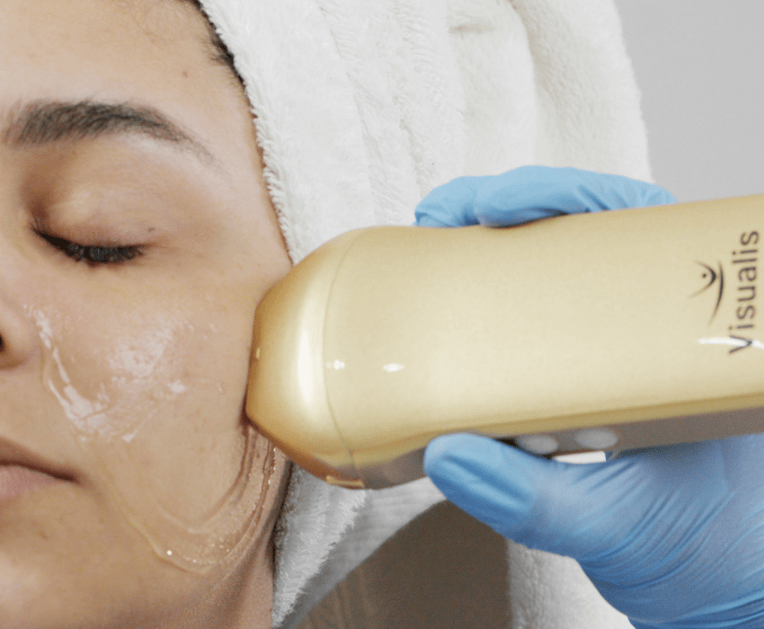
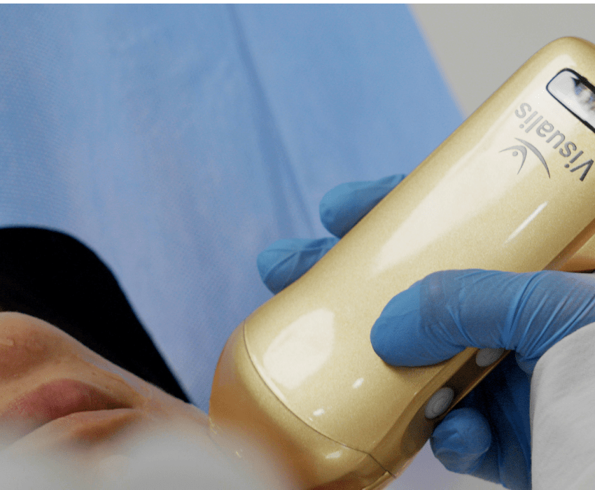
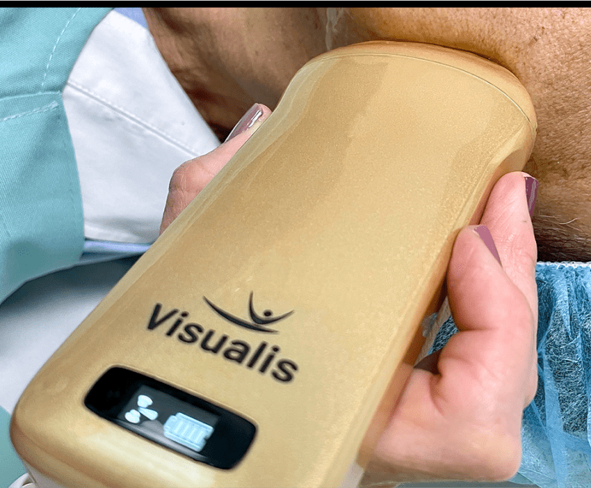
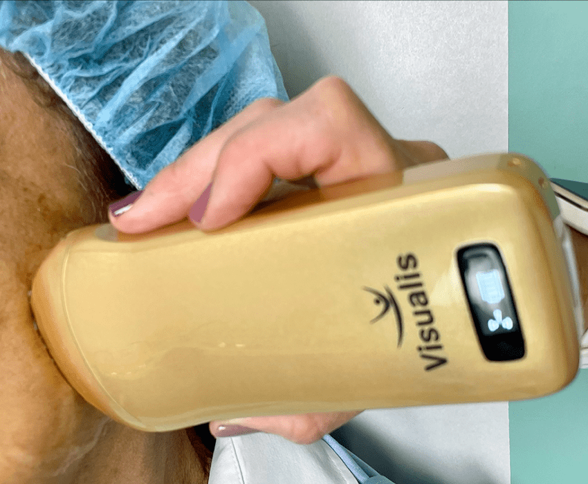
The Visualis portable ultrasound machine enhances your professional aesthetic practice, as it allows you to clearly visualize the superficial facial and body anatomy that is under the external layer in real time, thereby enabling you to make better decisions when determining the most appropriate aesthetic medical treatment for each patient, guiding cosmetic filling procedures with safety and treating complications with confidence.
Diagnose and guide your procedures
Map and inject safely
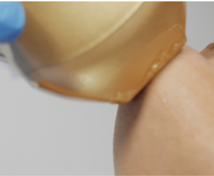
Visualize facial arteries in high-risk areas with high-definition ultrasound for safer injections.
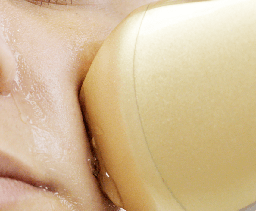
Use Visualis to accurately evaluate the location and depth of facial arteries. There are anatomic variations in the pathways of blood vessels in the face, which is why the use of an ultrasound in high-risk areas is of vital importance to ensure safe aesthetic practice.
Clear visualization of vessels allows for safer injections followed by images to confirm filler placement and healthy vascular flow.
Evaluate fillers
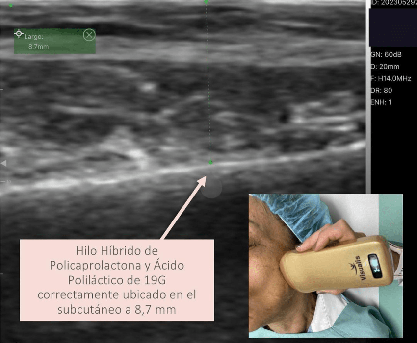
Confirm and characterize existing facial fillers using Visualis.
From confirming the placement of new fillers to diagnosing existing problematic ones, it is important to be able to confidently evaluate fillers by identifying their composition, location, and the health of adjacent tissue. The use of the high-definition ultrasound scanner Visualis allows for precise ultrasonic characterization to identify the types of fillers, document changes over time, and quickly confirm any complications.
The use of Visualis in aesthetic medicine is one more tool to show the patient the changes made from within their skin.
Dissolves fillers
Dissolves problematic fillers of hyaluronic acid and other compounds
Complications of fillers, such as swelling, migration, or vascular occlusions that can cause skin necrosis or blindness, can be treated with a smaller volume of hyaluronidase thanks to greater injection precision. Watch the injection in real-time on a nodule and see how the filler dissolves in front of you.
Studies that support the use of ultrasound in aesthetic medicine
Schelke et al. concluded that ultrasound examination can be an important tool to improve the safety of hyaluronic acid filler treatments.
In his study, González says that ultrasound is indicated in patients with complications derived from the use of different filling materials, when it is desired to discard the coexistence of multiple filling substances, establish their degree of absorption, and as a prior examination to the injection of a filling material in patients suspected of previous application of these substances and who deny, do not recognize, or do not remember their use.”
Alfageme et al. determined that the use of US is expanding in aesthetic medicine since US can provide relevant information that includes data on facial anatomical variants, the type, location and extent of common cosmetic fillers, the identification of implants, the complications of lipolytic procedures, and the possibility of percutaneous US guidance for the procedure.
In summary
Visualis allows you to have:
- Objective evaluation of aging.
- Identification of the patient's anatomy.
- Measure and locate the fat thickness before and after treatment.
- Objectification of results.
- Diagnosis of previous facial fillers.
- Choice of treatments.
- Study of complications.
- Treatment of complications.
- Ultrasound-guided treatments.
- Images for research work.
Other applications of Visualis
Our Visualis linear scanner also offers high definition images of superficial structures such as musculoskeletal system, eyes, nerves, blood vessels, thyroid, chest, breast and lungs, in both adult and pediatric patients.
Visualis is ideal for Aesthetic Medicine and Dermatology, but it is also useful in Plastic Surgery, Traumatology, Anesthesiology, Oncology, Ophthalmology and other specialties requiring images of superficial structures.
Learn how to use Visualis
X
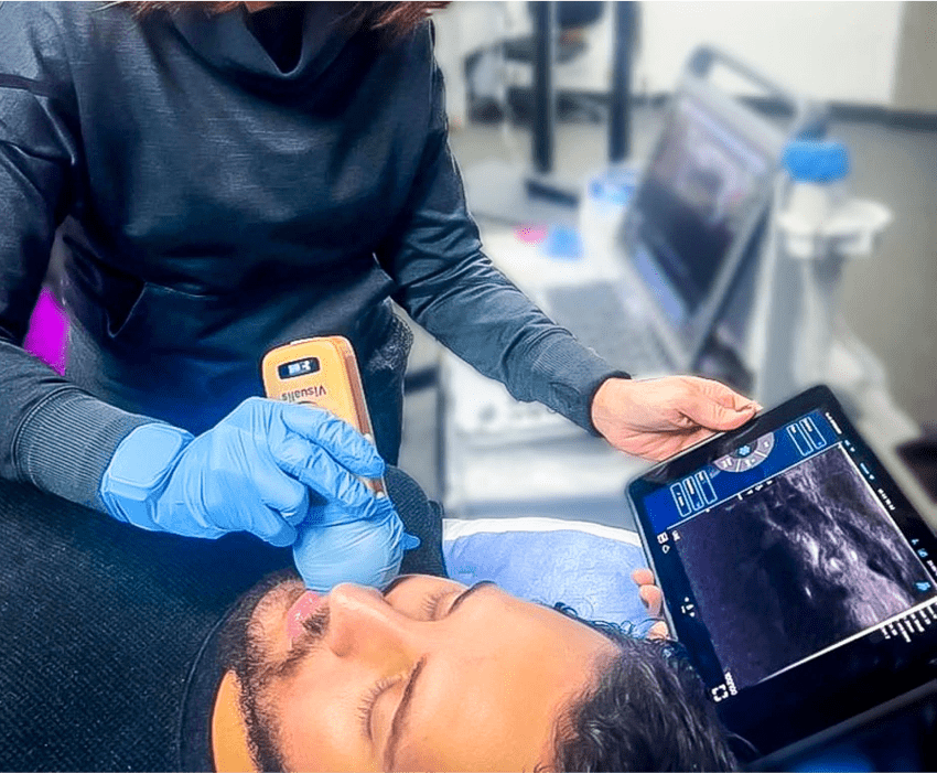
Visualis has changed the practice of aesthetic medicine in different aesthetic centers. Learn from Dr. Jorge León how to perform a soft tissue ultrasound of the facial region with Visualis, to recognize the facial artery and its branches.